Author: Nitya Jayaprakash
Abstract
Developing effective alternatives towards animal testing has been an area of study in recent years. Animal testing has been common in clinical trials
Introduction
Throughout human history, animals have been used for various human needs, including their role in scientific research and innovation. Animal testing has been important in advancing our understanding of biology, pharmacology, and medicine. (Hajar 2011) For example, pigs are increasingly used in studying traumatic brain injury (TBI) mechanisms due to their anatomical and physiological similarities to humans (Kinder, Baker, and West 2019). Their brain size, structure, and composition closely resemble those of humans, making them ideal models for understanding TBI. In chemical toxicity testing, animals have been used to assess the potential hazards of various substances (Chinedu, Arome, and Ameh 2013). An example of acute toxicity-LD testing involves administering a single dose of a substance to animals, usually rodents, to determine the lethal dose that causes death in 50% of the test subjects (Chinedu, Arome, and Ameh 2013) (Bruce 1985).
Observing the physiological and toxicological responses in animals can provide valuable information on how humans might react to the same substances. Animal models have been used to determine lethal doses, identify carcinogens, and evaluate the safety of cosmetic and industrial chemicals. (Löwa et al. 2018). Studies also show that animal tests predict human safety correctly only 50–70% of the time, highlighting a potential limitation of animal models being their significant biological differences from humans. Variations in genetics, metabolism, physiology, immune responses, and lifespan can affect the relevance of animal testing in predicting human reactions to chemicals.(Shanks et al. 2009).
However, the ethical concerns over the painful procedures and the suffering the animals endure leads to a call for change in these procedures. (Pacharinsak et al. 2022). The Institutional Animal Care and Use Committee (IACUC) is a regulatory body responsible for overseeing the ethical use of animals in research. It ensures that research involving animals is conducted humanely and follows established guidelines and regulations. The three R’s of animal testing- reduction, replacement, and refinement- gives us a guideline to improve the welfare of animals in experiments. Reducing the numbers of animals used, replacing animals with alternatives, and refining the procedures done to minimize suffering and pain, are guidelines followed in the industry, but we cannot accurately determine the pain, suffering, and distress experienced by the non-verbal animals. (Díaz et al. 2020)
While researchers are required to adhere to protocols that minimize pain, such as using anesthesia or euthanasia when appropriate, the nature of some tests inherently involves distress. Animal testing procedures can cause significant pain through injections, skin and eye irritation tests, and inhalation tests. A short-term study was conducted to evaluate the inhalation toxicity of nanomaterials using rats. The animals were exposed to aerosolized test materials at concentrations ranging from 0.5 to 50 mg/m³ over five consecutive days, followed by a 14- or 21-day observation period after exposure. Researchers analyzed bronchoalveolar lavage fluid (BALF) and examined sections of the entire respiratory tract. They also studied how the test materials were deposited in the lungs, cleared from the body, and whether they spread to organs outside the respiratory system. (Landsiedel et al. 2014).
This study highlights the potential for pain and distress in animal testing procedures. Prolonged exposure to varying concentrations of aerosols may cause respiratory irritation, inflammation, or damage, as evidenced by the need for BALF analysis and tissue examination. (Jirkof et al. 2010). The invasive methods used, including lung fluid extraction and histopathological assessments, highlights the physical harm inflicted on the animals. (Daston et al. 2015). Additionally, the uncertainty about the toxic effects and their systemic impact on the body likely hints at suffering and distress for the test subjects. (Landsiedel et al. 2014).
The exact number of animals experiencing pain and distress in the United States is difficult to determine properly because rats and mice, which are among the most commonly used species in toxicity testing, are not subject to the same reporting requirements. In 2000, 104,202 animals, accounting for 7.4% of all reported animals, were documented to have unrelieved pain and distress. (Stokes 2002).
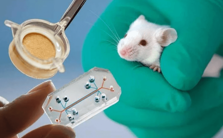
Efforts are made to develop alternative methods that reduce and/or eliminate animal suffering. Alternative methods to animal testing exist, however, it is unclear how effective these methods may be in comparison to animal testing. Therefore, in this review, we will highlight several alternative methods to animal models and discuss how effective each method is. (Doke and Dhawale 2015)
Cell Culture
In cosmetics, cell cultures are used for safety and efficacy testing, allergy testing, and examining anti-aging effects. It involves the growth of cells or tissue outside the body under controlled conditions. Research on tissue and organ formation, function, and pathology is possible largely due to the use of cell culture systems and animal models. They are frequently used in preclinical drug research, cancer studies, and investigations of gene function. (Foglietta et al. 2020). Animals are still used in cell cultures as sources for primary cells and tissues, particularly for studying complex systems that cannot be replicated. While many cell types can be cultured and immortalized, certain cells must be obtained directly from animals. (Arango et al. 2013)
Research indicated that solid tumors rapidly adapt to chemotherapy, with changes appearing soon after cellular exposure to drugs. Many factors influencing the effectiveness of anti-cancer drugs in vivo can be replicated in vitro using 3D cell culture models.(Duval et al. 2017) Immortal cell lines are invaluable in biomedical research, offering unlimited proliferation, consistency, and cost-effectiveness. They are used in applications such as regenerative medicine, drug delivery, bioproduction, disease modeling, and toxicology studies. By providing a renewable source of genetically modifiable cells, these lines facilitate efficient and reproducible experiments. (Voloshin et al. 2023)
Two-dimensional cell cultures have been instrumental in cancer research due to their simplicity, cost-effectiveness, and scalability. They provide a controlled environment for studying cellular behaviors and drug responses. (Abuwatfa et al. 2024)
2D and 3D cell cultures both represent research techniques with unique strengths. 2D cultures offer simplicity, cost-effectiveness, and rapid screening capabilities, while 3D cultures provide more physiologically relevant models with enhanced cell interactions and more accurate drug response predictions. (Jensen and Teng 2020). Though 2D systems are easier to manipulate, 3D models better mimic in vivo conditions. Each approach has its own uses and effectiveness in biomedical research. (Kapałczyńska et al. 2018).
Cell culture is scalable, cost-effective, and allows for quick, controlled experiments, but it doesn’t fully replicate how cells behave in a living organism. Animal testing helps study full body processes and is closer to human biology but has more costs, is more time-consuming, and raises ethical issues. Researchers may choose based on their goals and experiments, ideally with a shift towards using advanced cell culture techniques to reduce animal testing.(Balls 1994).While 3D cell cultures are improving in their ability to mimic tissue structures, animal models remain more useful for certain studies, particularly those involving complex biological interactions.
One of the pitfalls is that the 2D cell culture is usually homogenous – and to overcome this homogeneity, you can co-culture cells to simulate an in vivo environment while still maintaining the benefits of cost and ethics. (Antoni et al. 2015)
Organoids
Organoids are three-dimensional microsystems that come from stem cells that self-organize to mimic the structure, function, and physiology of intact organs, including their tissue-level activities and certain disease characteristics (van Berlo et al. 2021). One study generated midbrain organoids to model Parkinson’s disease to study the pathophysiology of the disease and provide a more accurate representation of human physiology than traditional cell cultures. In this study, researchers developed human midbrain organoids containing dopaminergic neurons, the cell type most affected in Parkinson’s disease. These organoids exhibited key features of midbrain development and function, including the production of dopamine and the presence of neuromelanin, a pigment found in human midbrain neurons. By introducing genetic mutations associated with Parkinson’s disease into these organoids, the researchers observed cellular phenotypes. The use of organoids can lead to a better understanding of disease mechanisms and the development of more effective therapies (Smits et al. 2019).
Reconstructed human skin models replicate human skin, providing an area for testing topical products. Tumor-specific stem cells can grow into 3D structures from small pieces of tumor placed in a synthetic matrix in a cell culture plate with various growth factors. (Lee and Koehler 2021)
These methods improve the safety and effectiveness of drugs, an example being that organoids are effective and safe models for studying lung diseases, demonstrated by their use in modeling SARS-CoV-2 infection and confirming alveolar cell marker expression through single-cell RNA sequencing. They offer a more realistic and complex environment than traditional 2D cultures, improving the predictability of pre-clinical studies. (Purev et al. 2024).
Organoids allow for the study of intricate interactions between different cell types. Liver organoids have been used to study drug metabolism and toxicity.(Lam et al. 2021). These 3D models, which can include one or multiple cell types, have diverse applications, from creating patient-specific liver cells to modeling chronic liver diseases and developing regenerative therapies. (Helke and Swindle 2013). The Liver Organoid-based Toxicity screen is a potential tool for liver toxicology studies, aiding in compound optimization, mechanistic research, precision medicine, and drug screening.(Palazzolo et al. 2022).While organoid technology bridges the gap between cell lines and in vivo models, the current system still has limitations. For example, lung organoids hold promise for tuberculosis research, but challenges remain before they can be fully utilized. Key issues include incorporating immune cells to better mimic immune responses and adding vasculature to create a more dynamic, controllable model, a problem nonexistent in animal models (Barkauskas et al. 2017).
Works Cited
https://onlinelibrary.wiley.com/doi/full/10.1111/exd.13498
https://link.springer.com/article/10.1186/1747-5341-4-2
https://www.sciencedirect.com/science/article/abs/pii/B9780128211809000052
https://link.springer.com/article/10.1186/1743-8977-11-16
https://academic.oup.com/ilarjournal/article/43/Suppl_1/S31/756764
https://link.springer.com/article/10.1007/s00204-014-1421-5
https://www.sciencedirect.com/science/article/pii/S2468202021000413
https://www.sciencedirect.com/science/article/pii/S1319016413001096#bb0040
https://www.nature.com/articles/s41531-019-0078-4.
https://www.sciencedirect.com/science/article/abs/pii/S0024320520305324
https://www.mdpi.com/1422-0067/24/16/12716
https://jbiomedsci.biomedcentral.com/articles/10.1186/s12929-024-00994-y
https://www.frontiersin.org/journals/neurology/articles/10.3389/fneur.2024.1378620/full
https://onlinelibrary.wiley.com/doi/full/10.1111/exd.14292
https://www.mdpi.com/2218-273X/14/1/115
https://journals.physiology.org/doi/full/10.1152/physiol.00036.2016
https://pmc.ncbi.nlm.nih.gov/articles/PMC6040128/
https://www.mdpi.com/2305-6304/10/2/91
https://journals.sagepub.com/doi/pdf/10.1258/002367794780681714
https://www.mdpi.com/1422-0067/16/3/5517
https://www.frontiersin.org/journals/molecular-biosciences/articles/10.3389/fmolb.2020.00033/full
https://www.frontiersin.org/journals/behavioral-neuroscience/articles/10.3389/fnbeh.2010.00165/full
https://www.ncbi.nlm.nih.gov/books/NBK459464/
https://link.springer.com/article/10.1186/s13619-021-00089-1
https://journals.lww.com/hrtv/fulltext/2011/12010/animal_testing_and_medicine.11.aspx
https://www.sciencedirect.com/science/article/abs/pii/0272059085900594
